Spectrum-Imaging
The software available on this page specializes in the treatment of STEM (Scanning Transmission Electron Microscopy) spectrum-imaging data using Multivariate Statistical Analysis (MSA), such as Principal Component Analysis (PCA).
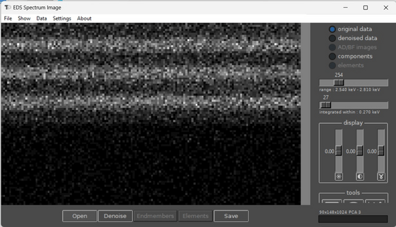
Spectrum Imager
Stand-alone tool for treatment of EELS and EDX spectrum-images.
Main Features:
>> Read most common formats of spectrum-imaging and convert data. Supported formats:
| Application | Extension | Read | Write |
|---|---|---|---|
| DigitalMicrograph (Ametek) | dm3, dm4 | 1D, 2D, 3D | 1D, 2D, 3D |
| Velox (ThermoFisher) | emd | 1D, 2D, 3D | No |
| PantaRhei (CEOS) | prz | 1D, 2D, 3D | 1D, 2D, 3D |
| ESPRIT (Bruker) | bcf | 3D | No |
| EDAX APEX (Ametek) | pts | 2D, 3D | No |
| Hyperspy 3D STEM | hspy | 1D, 2D, 3D | 1D, 2D, 3D |
| numpy | npy | 1D, 2D, 3D | 1D, 2D, 3D |
| binary | raw | 1D, 2D, 3D | No |
| Jeol STEM | pts | 2D, 3D | No |
>> Perform “silent” PCA denoising with adjustable number of components.
>> Cluster data in factor space with visualization of clusters and spectra.
Introduction to Spectrum Imager (PDF)
Download Learning Examples
MSA Plugin for DigitalMicrograph
Caution: debugging for new GMS versions is not supported anymore.
DigitalMicrograph plugin enabling PCA, ICA, Varimax rotation, and clustering of STEM EELS/EDX cubes.
last updated 3.08.2022, version 2.37
Application Examples
| Simulated Mg-Al-O (EELS) | Download (10.3 MB) |
| Si-Ge layers (EELS) | Download (37.4 MB) |
| Si-Ge layers (EDX) | Download (2.3 MB) |
| CMOS device (EDX) | Download (53.8 MB) |
| CMOS device (EELS) | Download (53.9 MB) |
| Superalloy (EDX) | Download (15.2 MB) |